Upper Back Anatomy Organs : Lower Back Muscle Anatomy And Low Back Pain / Cells, tissues, organs, organs systems and organs apparatus.. Diagram back muscles upper back human anatomy diagram anatomy. The deeper veins are buried well beneath the skin surface and run parallel to the arteries. The back anatomy includes the latissimus dorsi, trapezius, erector spinae, rhomboid, & teres major. Unit three — abdominal organs, pelvis & lower limb. How to draw the upper back anatomy and motion proko.
Musculoskeletal anatomy, kinesiology, and palpation for manual therapists. This article looks at the anatomy of the back, including bones, muscles, and nerves. The upper border articulates with the frontal bone and the anterior with the nasal; Integumentary, skeletal, muscular, nervous, endocrine, cardiov… a tendency to maintain a balanced or constant internal state This middle section of the small intestine has extensive folds to facilitate both secretion of digestive.
Tissues And Organs Fundamentals Merck Manuals Consumer Version from www.merckmanuals.com 14 photos of the upper back human anatomy diagram. Spine anatomy mayfield brain spine cincinnati. The standard position in which the body is the standard anatomical position is agreed upon by the international medical community. Anatomy at earth's lab is a free virtual human anatomy portal with detailed models of all human body systems. Many conditions and injuries can affect the back. Palmar region , arteries (illustrations: Topographically, the muscles in this group are classed along with the lateral torso wall and upper extremity, which is due to their location as well as their genetic development based on their embryological origin. The deeper veins are buried well beneath the skin surface and run parallel to the arteries.
Cells, tissues, organs, organs systems and organs apparatus.
It also covers some common conditions and injuries that can affect the. The human back, also called the dorsum, is the large posterior area of the human body, rising from the top of the buttocks to the back of the neck. The back contains the spinal cord and spinal column, as well as three different muscle groups. These autonomic components conduct the unconscious signals that control the organs and glands of the body. Unit three — abdominal organs, pelvis & lower limb. 14 photos of the upper back human anatomy diagram. Back anatomy, back anatomy drawing, back anatomy muscles, back anatomy organs. Click on the labels below to find out more about your organs. Topographically, the muscles in this group are classed along with the lateral torso wall and upper extremity, which is due to their location as well as their genetic development based on their embryological origin. How to draw the upper back anatomy and motion proko. Learn about back anatomy physiology upper with free interactive flashcards. Assessment | biopsychology | comparative | cognitive | developmental | language | individual differences | personality | philosophy | social | methods | statistics | clinical | educational | industrial | professional items | world psychology |. Wolters kluwer health/lippincott anatomy and human movement:
They originate from the vertebrae and insert into the scapulae. Organs exist in most multicellular organisms, including not only humans and other animals but also plants. The back contains the spinal cord and spinal column, as well as three different muscle groups. Chemical, cellular, tissue, organ, system, organism. Includes the study of the gross and microscopic structure of the systems of the human body with special integrates anatomy and physiology of cells, tissues, organs, the systems of the human body, and mechanisms responsible for homeostasis.
Levator Scapulae Muscles In Isolation Rear View Of Upper Back Human Anatomy Stock Illustration Adobe Stock from as1.ftcdn.net Superficial veins of upper limb , anatomy : Nervous system, skeleton, front view of muscles, back view of muscles. Integumentary, skeletal, muscular, nervous, endocrine, cardiov… a tendency to maintain a balanced or constant internal state Their main function is exchanging oxygen and. The human back, also called the dorsum, is the large posterior area of the human body, rising from the top of the buttocks to the back of the neck. Topographically, the muscles in this group are classed along with the lateral torso wall and upper extremity, which is due to their location as well as their genetic development based on their embryological origin. Structure and function (6th ed.). The cause may be poor posture (such as forward head posture) or any type of irritation of the large back and shoulder muscles, including muscle strain or spasms.
Learn about back anatomy physiology upper with free interactive flashcards.
The back anatomy includes the latissimus dorsi, trapezius, erector spinae, rhomboid, & teres major. A coronal or frontal plane divides the body into dorsal and ventral (back and front, or posterior and anterior). Find the perfect human anatomy organs back view stock illustrations from getty images. There is a vast tubular network of veins just below the skin of the upper extremity. The two lungs are located on either side of the upper chest. The cause may be poor posture (such as forward head posture) or any type of irritation of the large back and shoulder muscles, including muscle strain or spasms. Many autonomic nerves and ganglia pass through the thoracic region to innervate the internal organs. How to draw the upper back anatomy and motion proko. Click on the labels below to find out more about your organs. It is very stiff, and the thoracic spine has a limited range of motion. Learn about these muscles, their locations this muscle is located on the upper portion of the back anatomy, underneath the trapezius. Organs exist in most multicellular organisms, including not only humans and other animals but also plants. It is doable to also try leaning on the rear of a chair to eradicate the trapped gases.
Back muscle diagrams labeled diagram anatomy organ charter. Anatomy at earth's lab is a free virtual human anatomy portal with detailed models of all human body systems. Click on the labels below to find out more about your organs. Bones of the upper limb | anatomy and physiology. They originate from the vertebrae and insert into the scapulae.
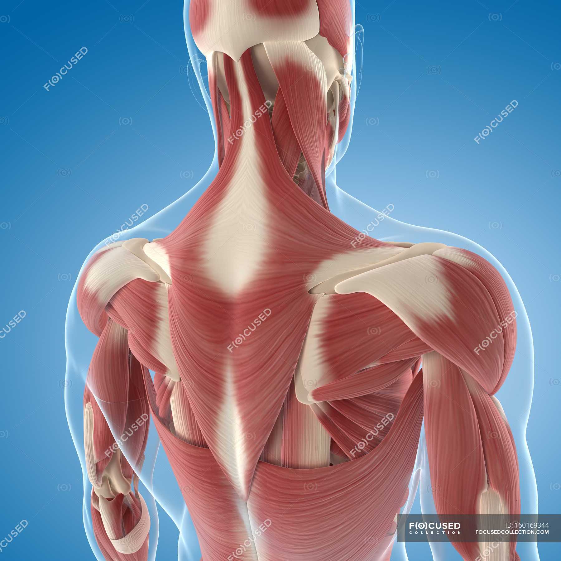
Upper Back Musculature Muscle Groups Anatomy Stock Photo 160169344 from st.focusedcollection.com The extrinsic back muscles are also referred to as secondary back muscles. Superficial veins of upper limb , anatomy : The standard position in which the body is the standard anatomical position is agreed upon by the international medical community. Includes the study of the gross and microscopic structure of the systems of the human body with special integrates anatomy and physiology of cells, tissues, organs, the systems of the human body, and mechanisms responsible for homeostasis. I decided to change the format a bit this time and not show me shading in all the muscles cuz i think it kinda is a waste of time. We look at why mobility is so the nerves that supply all of the internal organs emerge from the thoracic vertebrae, so it has quite a significant responsibility to deliver its goods! The upper border articulates with the frontal bone and the anterior with the nasal; The deeper veins are buried well beneath the skin surface and run parallel to the arteries.
Bones of the upper limb | anatomy and physiology.
Anatomical diagram showing a front view of organs in the human body. We look at why mobility is so the nerves that supply all of the internal organs emerge from the thoracic vertebrae, so it has quite a significant responsibility to deliver its goods! Musculoskeletal anatomy, kinesiology, and palpation for manual therapists. This article looks at the anatomy of the back, including bones, muscles, and nerves. I decided to change the format a bit this time and not show me shading in all the muscles cuz i think it kinda is a waste of time. Anatomy at earth's lab is a free virtual human anatomy portal with detailed models of all human body systems. Upper back pain is most commonly caused by muscle irritation or tension, also called myofascial pain. The human back, also called the dorsum, is the large posterior area of the human body, rising from the top of the buttocks to the back of the neck. Diagram back muscles upper back human anatomy diagram anatomy. Many autonomic nerves and ganglia pass through the thoracic region to innervate the internal organs. 14 photos of the upper back human anatomy diagram. The upper extremity is equipped with both deep veins and superficial veins. The upper limb is the organ of the body, responsible for manual activities.
Development of the human organism upper back anatomy. Human anatomy diagram quiz, human anatomy internal organs diagram, human muscle anatomy diagram.


0 Komentar